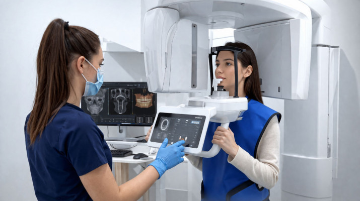
Cone Beam Computed Tomography (CBCT) has transformed modern dental diagnostics, offering clinicians unprecedented insight into patient anatomy at a relatively low radiation dose.
What Is CBCT?
CBCT (Cone Beam Computed Tomography) is a three-dimensional imaging technique developed specifically for maxillofacial structures. Unlike conventional computed tomography (CT), which uses a fan-shaped X-ray beam, CBCT employs a cone-shaped beam that rotates around the patient’s head and records images using a flat-panel digital detector.
The examination produces a volumetric dataset – a so-called voxel grid (three-dimensional pixels) – enabling reconstruction and analysis of structures in any plane: axial, coronal, and sagittal, as well as in 3D projections.
A Brief History of the Technology
The first CBCT devices dedicated to dentistry appeared at the turn of the 21st century. The NewTom QR DVT 9000, designed by Italian engineers and introduced to the market in 1999, is regarded as one of the pioneering systems. Since then, the technology has developed rapidly – exposure times have shortened, detector resolution has improved, and diagnostic software has gained advanced treatment-planning tools.
How Does CBCT Work?
The CBCT image acquisition process proceeds as follows:
- Patient positioning – the patient sits or stands still (depending on the device model), with the head stabilised in the unit’s positioning frame.
- Arm rotation – the radiation source and detector rotate synchronously around the head through an arc of 180°–360°, capturing anywhere from several dozen to several hundred projection images.
- Reconstruction – specialised software processes the projection images using filtered back projection or iterative algorithms to create a three-dimensional volumetric model.
- Analysis – the clinician reviews the data in dedicated software (e.g. Romexis, i-CAT Vision, Planmeca Romexis), freely sectioning the volume and measuring anatomical structures.
Clinical Applications
1. Implantology
CBCT is considered the gold standard in implant treatment planning. It enables precise assessment of:
- bone height and width of the alveolar process at the planned implant site,
- topography of anatomical structures – the mandibular canal, maxillary sinuses, and mental foramen,
- bone density (Hounsfield scale), which may influence implant selection and the surgical protocol,
- the possibility of bone augmentation and selection of the optimal reconstructive technique.
3D planning software (e.g. coDiagnostiX, Simplant, Nobel Clinician) enables virtual implant placement and the production of a 3D-printed surgical guide, improving procedural precision and patient safety.
2. Endodontics
In root canal treatment, CBCT provides information unavailable from conventional two-dimensional radiography:
- root canal system morphology – additional canals (e.g. MB2 in maxillary molars), C-shaped canals, and anastomoses,
- precise working length and canal curvature,
- root perforations, internal and external resorption,
- inflammatory periapical lesions – their actual extent and relationship to neighbouring structures,
- causes of treatment failure – fractured instruments, blocked canals, and missed canals.
Clinical studies confirm that CBCT can increase the detection rate of periapical lesions compared with periapical radiography by as much as 35–50%, particularly in early stages.
3. Orthodontics
In orthodontic and orthognathic treatment planning, CBCT enables:
- three-dimensional cephalometric analysis – skeletal measurements in 3D space,
- assessment of the position of tooth germs and ectopic impacted teeth (particularly maxillary canines),
- analysis of the temporomandibular joints (TMJ) – including condylar shape and joint space,
- virtual planning of orthognathic procedures and simulation of treatment outcomes,
- diagnosis of dentoalveolar and skeletal abnormalities in three planes.
The integration of CBCT data with optical scans of the dental arches (intraoral scanners) is becoming increasingly common, creating a complete digital model of the patient.
4. Oral and Maxillofacial Surgery
Before procedures involving the craniofacial region, CBCT enables the surgeon to:
- accurately assess impacted teeth and their relationship to adjacent structures (e.g. neighbouring tooth roots and the mandibular canal),
- plan the removal of jaw cysts and bone tumours,
- diagnose and treat craniofacial fractures,
- assess paranasal sinus pathologies in the context of sinus lift procedures,
- virtually plan reconstructive surgery.
5. Periodontology
In the treatment of periodontal disease, CBCT provides precise information on:
- the depth and configuration of bone defects (vertical, horizontal, and furcation defects),
- the thickness of the cortical plate and morphology of the alveolar bone,
- the relationship between periodontal defects and root canals (endo-perio differential diagnosis),
- the three-dimensional extent of inflammatory processes.

CBCT vs Conventional Radiography – Comparison
The fundamental difference between the two methods lies in image dimensionality: conventional 2D X-rays provide a flat image, whereas CBCT generates a complete three-dimensional reconstruction. Examination times are comparable – a panoramic radiograph typically takes 10–20 seconds, while CBCT takes 5–40 seconds depending on the device and selected field of view.
In terms of radiation dose, 2D radiography remains the lower-dose method, while CBCT involves moderate exposure (although still significantly lower than conventional CT). The same applies to cost: X-ray examinations are considerably less expensive, and panoramic and periapical systems are widely available; CBCT is more costly, although access to the technology continues to expand.
Both methods share limited soft-tissue resolution – 2D radiography and CBCT are primarily used to assess bone and dental structures and do not replace magnetic resonance imaging for soft-tissue imaging. The key difference concerns clinical indications: 2D X-rays are highly effective for screening and routine monitoring, whereas CBCT is a tool for complex diagnostics and detailed planning of surgical, implantological, and endodontic procedures.
Radiation Dose and Safety
One of the key considerations is exposure to X-ray radiation. CBCT emits a significantly lower dose than conventional head computed tomography (CT), but a higher dose than standard dental X-rays.
Approximate Effective Radiation Dose Values:
- Periapical X-ray (1 image): ~1–8 µSv
- Panoramic radiograph: ~5–22 µSv
- CBCT (small field of view – FOV): ~30–100 µSv
- CBCT (large field of view – FOV): ~100–600 µSv
- Head CT: ~1000–2000 µSv
- Background radiation (annually): ~2000–3000 µSv
For comparison, a single CBCT examination corresponds approximately to several hours of transatlantic air travel.
The ALARA Principle
Dentistry follows the ALARA principle (As Low As Reasonably Achievable) – the radiation dose should be minimised while maintaining the diagnostic value of the image. In practice, this means:
- selecting the smallest field of view (FOV) covering only the clinical area of interest,
- optimising exposure parameters (kV, mA, time),
- using lead shielding (apron, thyroid collar),
- referring patients for the examination only when the results are expected to influence the treatment plan.
Technical Parameters of CBCT Devices
Field of View (FOV)
FOV determines the anatomical volume covered by the examination:
- Small FOV (< 10 cm): examination of a limited area, e.g. a single quadrant – endodontics, implant diagnostics. Lowest radiation dose.
- Medium FOV (10–15 cm): full dental arch or both jaws.
- Large FOV (> 15 cm): the entire craniofacial complex – orthognathic procedures, orthodontics, surgery.
Isotropic Resolution (Voxel Size)
The smaller the voxel, the higher the image resolution. Typical values include:
- 0.08–0.15 mm – high resolution, used in endodontics and implantology,
- 0.2–0.4 mm – standard resolution in orthodontics and surgical planning.
Acquisition Time
Modern devices capture images in 5–20 seconds, minimising motion artefacts and improving patient comfort.
Integration with Digital Technologies
CBCT forms the foundation of the digital workflow in modern dental practice.
CBCT data are exported in DICOM (Digital Imaging and Communications in Medicine) format, which is compatible with planning software and enables information exchange between specialists.
Limitations of the Technology
Despite its many advantages, CBCT has certain limitations that clinicians should bear in mind:
- Metal artefacts – amalgam fillings, crowns, and implants can cause streaking, shadowing, and bright artefacts that hinder interpretation. Newer metal artefact reduction (MAR) algorithms partially reduce this problem.
- Low soft-tissue resolution – CBCT does not adequately visualise soft tissues (mucosa, nerves, blood vessels), unlike magnetic resonance imaging (MRI).
- Patient immobility is required – movement during exposure degrades image quality.
- Interpretation requires specialised training – 3D images are diagnostically more complex than conventional X-rays; incorrect interpretation may lead to clinical errors.
- Equipment cost – CBCT systems are significantly more expensive than conventional panoramic units.
Legal and Ethical Aspects
In Poland, CBCT examinations are subject to the provisions of the Atomic Law and regulations issued by the Minister of Health concerning medical exposure. Key principles include:
- Justification of the examination – every radiographic examination must be clinically justified; the referring clinician is responsible for the decision.
- Dose optimisation – techniques that minimise exposure must be used.
- Examination reporting – CBCT results should be interpreted and reported by an authorised physician (a radiologist or dentist with appropriate training).
- Protection of particularly vulnerable individuals – indications for CBCT in children, pregnant women, and oncology patients should be assessed with particular care.
The Future of CBCT in Dentistry
CBCT technology continues to evolve dynamically. Key directions of development include:
- Artificial intelligence (AI) in image analysis – algorithms supporting the identification of pathological changes, automatic anatomical measurements, and segmentation of structures (e.g. nerves and sinuses).
- Dose reduction while maintaining image quality – new detectors and iterative reconstruction algorithms make it possible to obtain diagnostically useful images at increasingly lower exposure levels.
- Integration with surgical robotics – CBCT-based intraoperative navigation systems (intraoperative CBCT) enable real-time verification of implant position.
- Combination of CBCT with MRI – image fusion techniques combine the high bone-detail accuracy of CBCT with the soft-tissue resolution of MRI.
- Portable CBCT devices – miniaturisation of the technology opens up possibilities for bedside diagnostics and use in settings with limited access to conventional imaging infrastructure.
Summary
CBCT is one of the most transformative technologies to enter modern dentistry. Three-dimensional imaging of craniofacial structures has revolutionised diagnostics and treatment planning in implantology, endodontics, orthodontics, and oral and maxillofacial surgery. With appropriate clinical indications, a properly selected field of view, and adherence to the ALARA principle, the benefits to the patient can significantly outweigh the risks associated with radiation exposure.
The dentist of the future is a clinician who works confidently in a digital environment – integrating CBCT data with other diagnostic modalities and CAD/CAM technologies to plan treatment with surgical precision even before the procedure begins.